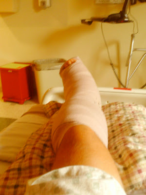
Monday, December 29, 2008

7 Weeks Post-Op ... depressed

It's been a while since I've been into the doctor's office. My mother-in-law passed away the week of Christmas and my appointments were postponed. I should have been in for a follow up 2 weeks ago, but things haven't worked out.

It's been icy this winter. I've stayed home when it's been bad. I'm lucky that I can do some work from home. I tried to go into work once, but had a bad slip on the ice and put all my weight on my left (healing) foot. It's a strange feeling. My whole leg stiffened involuntarily before I fell on it. I suppose to protect itself. It ached afterwords, but I am sure I haven't done any damage. I have fallen this way twice now. Once on the ice, and once in my kitchen on a stray paper towel. You can never be too careful.

I'm writing tonight because I cannot sleep and just had a bad run of diarrhea. I stopped taking Vicadin two days ago and have just been taking Tylenol. I can't help but think that some of my symptoms are mild withdrawal symptoms. I have been taking Vicadin steadily for over a month.

Pain comes and goes. I mostly feel pain on my commute back and forth to work. I'm in the car for about 50 minutes in the morning and about an hour in the evening. My foot is down the whole time. The bumps in the road and the mild rocking back and forth causes some bone discomfort. The nerve pain mostly occurs at night when my foot has been in the same position for a while.

I keep my foot up whenever I can. It's still the best pain relief. But I am restless. Depression has really been a big struggle. We have three children; all boys. During the holidays, the funeral and preparations, my wife still had to take care of us all. I had very little to offer in the way of help. It really stinks to be layed up like this. Was it worth it? Time will only tell.

I'm still wearing the boot and I'm still on crutches. I hate the boot. I'm sure anyone who has worn the boot also hates the boot. I'll sign this post off with a photo of my least favorite friend...the boot, and a Christmas Tree.

Friday, December 12, 2008

Scar Tissue Nerve Hypersensitivity

I'm four weeks post surgery now. I went to the doctor today because I'm still having some bleeding and have been having some pain.

The pain I'm having is right at my incision points. The pain can be severe sometimes, and I liken it to a red hot poker or a red hot iron being pressed into the skin.

The burning pain is actually common. It's from scar tissue nerve hypersensitivity. Here's what happens:

When the skin is cut, especially deeply as in surgery, the nerves that cross the incision are severed. As they heal, new nerve endings are created. These nerve endings have never experienced the world before so anything touching them; a sock, cast, boot etc. causes them to go off like fireworks. And it hurts.

The fix is to take vitamin E oil and rub it into the scar – rub it HARD. And this, of course, hurts like heck. But the idea is to desensitize the area.

I'll let you know how it goes in a couple of weeks.

A little update:
The vitamin E oil treatment works. The oil doesn't actually help from a medicinal point of view — it's really just a lubricant. But it does make the scar look a ton healthier. I'm at eight weeks now and occasionally the scar on the top of my foot will ache. If I rub it, the pain goes away. Sweet.

Sunday, November 23, 2008

Follow up #2

At my second follow up the cast came off and they took X-rays. I'll post them as soon as I can.

Everything seems to be healing well, and the staples and sutures came out. This did hurt a bit, kind of a burning sensation.

They put a tube sock on my foot and gave me a boot. I can't bend my foot enough to get it all the way in, I'm suppose to work at it though.

This particular boot has a a little push pump on it that airs everything up tight after I get it on.

I'll be able to shower without bagging up a cast. I can take the boot off. Yeah!

UPDATE...Here's the X-rays (click to enlarge):

I've duplicated one x-ray here and highlighted areas of interest. The little white "C's" up and down the back of my leg are the staples before they were removed. I'm still not weight-bearing. I'll have new x-rays then.


This is the money shot of the screw in my heel bone. Imagine sitting on the floor at the foot of my recliner and looking up through the arch of my foot at an angle. That's the angle of this x-ray.

Wednesday, November 19, 2008

Follow up #1

Warning! This article features graphic post-operative surgery photos.

I had my first follow-up today. One week after my surgery. I was tense as the nurse began to unwrap my leg. I hadn't seen it in a week and had no idea what I was about to see. I had felt little tingles, itches and minor pain here and there, so I had visualized in my mind what things would look like. The gauze had a lot of dried blood in it, which was a little gross. But no fresh blood was there. I left the office with cleaned wounds and a brand new orange cast. I'll have the cast for one week. Now, I apologize for the photo quality. My camera was broken, so I had to borrow my son's. Here are the pictures with a little commentary:

^ Okay, this was a little incision in my shin right below my knee that I wasn't expecting. This incision had one staple in it, which was removed. What the surgeon did here was harvest some bone marrow from the thickest part of my tibia. He mixed this marrow with the cadaverous bone graft material to create a graft that contained my own cells, thereby further encouraging bone growth.


^ This little beauty is the incision for the Cotton Osteotomy. This is the procedure that will push my big toe back down to recreate my arch. This is the first time I've ever seen an arch on my left foot. The line above the incision is just a drawing mark. The blue color around the base of my toes is where blood settled after the surgery. It looks like a bruise. This incision hurt a little bit after surgery.


^ This is the big one. This is the "outside" or lateral view of my left foot. This incision was where the talo-calcaneal coalition was broken, reset and packed with bone graft. This is also where the lateral column lengthening was done, whereby the calcaneous was elongated to straighten my foot. Again, the blue coloring below the incision is where blood settled giving a bruised appearance. This incision doesn't hurt much, but I feel a lot of pressure here whenever I stand up on my crutches. It goes away when I elevate it.


^ This is looking up at my heel. This incision gave the surgeon access to the bottom of my heel bone where he placed the screw up through the heel bone into the talus. This incision gets a little sore when my foot has set in one place for a while. You can see another incision in the lower left corner. I'll explain it next.


^ Okay, this is the last one. This is a shot of my calf. The wrinkly places are just impressions from the old gauze. The incision gave the surgeon access to the Achilles tendon, just below the calf muscle. The surgeon made a small incision into the tendon, which relaxes the tension on it. When the heel bone is repositioned it can pull the tendon tight. This helps relieve that.

Friday, November 14, 2008

Post-op: Part 2

I spent the night at the hospital. As the effects of the nerve block wore off I had a PAC machine hooked up to my IV to help with pain. The PAC machine has a big syringe inside of it full of pain killer. I was told that if I felt even a tingle I should hit the button that activated the PAC. The PAC would not allow me to overdose, so I could hit the button whenever I felt something.


While I was in the hospital I felt NO pain. Nothing. This was a real surprise for me as I was under the assumption that pain was going to be a real bear after the surgery.

We did keep ice on my foot to help with any swelling. I spent most of my time in the hospital watching TV and sleeping. I was discharged less than 24 hours after surgery.

Getting around the house
A physical therapist trained me on how to use crutches, which was a little scary. I have a friend in the medical supply business that brought me a walker. It's really great and I would recommend you get one for around the house. It comes in especially handy in the bathroom when you need the extra stability.

Bathing
I'm not allowed to shower or take a bath for a week. This is a bit tough. I've been using a wash rag and my wife washes my hair.

Pain Medicine
I'm taking two Percocet 7.5/325mg every 5 hours. The directions say every 6 hours, but I start hurting after 4. So I split the difference. I am hurting now, but not horribly. I compare the pain I'm feeling to the way my foot felt after I spent the day walking or cutting the grass. I had the surgery because my foot hurt, so I am used to foot pain. But as the Percocet wears off, I do get sharp pains and some periods of throbbing. It's all manageable though. Keeping my foot elevated helps eliminate pain as well.

Follow-up
I will have a follow-up appointment with the PA (physician assistant) in about a week. I'll take my camera with me in case the bandages come off. I think I have a total of 5 incisions.

Thursday, November 13, 2008

Post-op: Part 1

It's been two days since my surgery and I'm doing fine. In this first post-op entry, I'm actually going to focus on my pre-op.

I arrived at the hospital two hours before the surgery was scheduled. I spent very little time in the waiting area (thank goodness), then my wife and I were brought to the pre-op room. This session began with several questions regarding the condition, getting my medical history up-to-date and confirming other information. They showed me a little gizmo that is used to encourage me to take deep breaths. I was to use it after the operation to help avoid pneumonia. They also started an IV.

Soon the physician assistant arrived as well as the anesthesiologist. The said they were getting ready to administer a block on my leg. Have you ever received a shot at the dentist for say, a filling? The dentist deadens a key nerve that numbs your whole face. In the case of your foot, the nerve is behind the knee. They rolled me over on my belly. Then a nurse put some medicine in my IV that she called a "six-pack." I don't know if I felt like I had just drank a six-pack, but it really relaxed me. I felt some tingling on my leg as they found the nerve and the shot really did not hurt too much. They rolled me back over and I think I received another shot in my shin. Forgive me, by this time I was feeling pretty out of it. My wife was not able to be in the room with me during this, so I don't have her to help me.

Gradually my foot fell asleep. It would stay that way for nearly 20 hours. They rolled me down the hall into the operating room and I don't remember much after that. Next thing I knew I was waking up in post-op. I felt a little groggy and my foot was still numb. The only thing that changed was now I had a big splint and wrap on my foot. Here's what I saw when I woke up, minus the ice bags:

Friday, November 7, 2008

CT Scans

My surgery is 5 days away now. The surgeon asked that I get a copy of my CT scan on a disk and bring it in before the surgery. So I have the disk, and I did a little search online for some CT scan program and found one. I downloaded the free trial version of an app called Osirix. (I use a Mac, BTW, and still found software.) I fired up the program and after messing with it a bit, I was able to grab a couple of shots of the coalition. Here's the first:

You can click the image to enlarge it. This is looking from the "front" of my left foot. The top bone is the bottom of my tibia. The little oval on the right is the bottom of the fibula. The chunk in the middle is the talus and calcaneous. I'm not a radiologist, so I didn't put any arrows on this one, it looks pretty fused to me. Here's another:


This one looks a bit more like a foot. My guess is that the bright white spot in the ankle is the fusion. I'll print these out and ask my doctor about it.

Okay, here's the fun part. The program I downloaded can take the CT scan data and build a 3D model. I can spin it around and mess with the contrast. As I adjust the contrast it gradually removes the skin and muscles and veins. Here's three views of MY foot. Freaky!


You can really see the talar beaking on this last image. I'll let you know how the surgery goes!

Monday, October 27, 2008

Surgery scheduled

Note: this post is updated regularly as I learn more.

I met with a orthopedic surgeon today who specializes in foot and ankle surgery. For the first time I had a doctor talk to me about other procedures that need to be done besides just the subtalar fusion that had always been recommended to me.

One of my main concerns about the fusion surgery was that it may not correct the position of my heel. Doctors had always said something like "We'll just complete the fusion." This never sounded "corrective" to me.

It was explained to me today that, if it is possible, the coalition will be broken apart and the heel bone will be moved to a more optimal position before fusion. That made me feel a ton better. Hopefully that will prevent the twisting sensation I feel when I walk. If they cannot move the calcaneous (heel) bone, it will need to be cut in half and repositioned (a calcaneal osteotomy). Apparently there are lots of nerves and arteries under the subtalar joint that limit how far you can move the calcaneous. My surgeon referred to this as an area of "major badness." I'll take his word for it.

But there are some other things the surgeon will do. Apparently my forefoot has compensated for my heel being misaligned. Once the heel is repositioned, my forefoot will be twisted (supinated) which may require more surgery. In order to compensate, the surgeon is recommending a lateral column lengthening (involving the calcaneous bone) and also a Cotton Osteotomy on a cuneiform. These involve wedging a bone graft into strategic areas to move things into the proper position. The achilles tendon will receive a small incision below my calf muscle. This releases some tension here to compensate for any tightening resulting from the heel mocement.. This will all be done at the same time as the subtalar fusion. I think I counted a total of 4-6 separate incision points.

The subtalar fusion will be the same as you read about everywhere else. Bone grafts and screws. The doctor said I will be non-weight bearing (crutches) for at least six weeks. After that I will gradually start to put weight back on the foot.


Click image to enlarge.

If you have tarsal coalition and are considering surgery, try to find a foot and ankle specialist. I have been to a few orthopedic surgeons and they each told me they had done the surgery a "few" times, which scared me a little. When I asked the specialist today how familiar he was with this surgery, he said he makes these type of corrections "all the time." So don't be afraid to quiz your doctors.

Radiology

I was able to get a copy of my X-rays from my podiatrist. I've posted here, as well as the report from my CT scan. I've noted a few things of interest on the X-rays. First is what is called the C-sign (white arrows), named because of the shape of the formation. I'm still researching what causes this. Next is the area of the coalition, which in my case is the middle facet between the talus and the calcaneous (red arrow). This is hard to see. Third is what is called beaking (because it looks like a beak). It's visible between the talus and the navicular (yellow arrow). Talar beaking is common with subtalar coalition. I'm looking into what causes beaking and if it's a source of pain. It sure looks like it is. Also, it's worth noting how much shorter the talus is in the left (coalition) foot than in the right.

If you're really interested, here is the radiology report from my left foot CT scan:
There is talocalcaneal coalition at the sustentaculum tali / middle talocalcaneal facet. The posterior talocalcaneal facet and anterior articulation appear preserved. There is dorsal talar beaking, foreshortening of the anterior talar process and dorsal navicular beaking all in a pattern typical for subtalar coalition. The ankle mortise is intact. There is foreshortening of the medial malleolus, also typical for subtalar coalition. The calcaneal cuboid joint, navicular cuneiform joints and all three intercuneiform joints are unremarkable. The Lis Franc joints are normal in appearance. The remaining osseous structures are unremarkable.

Flat feet

In “Why does it hurt?” I illustrated how the coalition causes the arch of my foot to collapse. I wanted to include one more picture. This is my “Charlie Chaplin” photo. I don’t actually stand like this, it’s two pictures put together.


The white lines show the distance from where my heel touches the ground to where the top of my arch is. I’ve also included a redline to show the difference between my ankles. This can cause problems all the way up the leg. I have tired legs and frequent back trouble. It’s either from this or I’m just getting old.

Now it’s important to know that there are two types of flat feet: flexible and rigid. Flexible flat feet can be treated with orthotics and inserts. These move the foot into the proper arch.

I’ve tried two kinds of orthotics for my feet. I hated them. I walked off the side of my shoes and they were painful. It is common to try orthotics for flat feet from tarsal coalition. And I’ll leave it to the experts to decide whether or not use them. But when you think about it, how does an orthotic help a rigid flat foot?

My orthotics twisted my leg until I had a condition in my knees called sublux patella. It had to be corrected with surgery. If your doctor is recommending this, quiz him or her about it. Sometimes orthotics and special shoes are not covered by health insurance so be careful what you spend your money on.

Why does it hurt?

When functioning properly, your foot forms a tripod, which is a stable foundation for your body. As you take a step, your heel actually moves from side to side. It moves to where it makes the tripod most stable.

When your heel has moved to the pinky toe side of the foot it is said to be in the valgus position. When it has moved to the big toe side it is said to be in the varus position.

In a foot that involves a coalition of the calcaneous (heel bone) with another bone, it is locked into the valgus position. This makes the tripod off center and the arch of the foot collapses. If you have a coalition, you have a flat foot.


In this image, I attempt to illustrate the tripod concept. Remember, my right foot is normal. When someone looks at the image on the left, they may think that the feet are completely normal. But if you look closely, you can see the heel of my right (normal) foot, but not my left (coalition) foot. You can also begin to see how my left (coalition) foot is "rolling" inward which makes my arch disappear. On the right I've drawn some tripods. You can see how the tripod of my right (normal) foot is stable. However, the the heel of my left foot is stuck in the valgus position which causes the point of the tripod to buckle (red arrow).

This instability causes all kinds of trouble. Tendons and other tissues get pulled and twisted abnormally which can cause a burning sensation on the top/outside of your foot. The muscle and tissue along the arch of your foot gets stretched and can spasm. The joint itself takes on a lot more stress than it's intended to.

You may also have some very deep pain, I know I do. That little bony bridge, the coalition, gets a ton of torque on it. The twisting and torquing can cause an bone marrow edema (collection of fluid) to form inside the bones themselves. A recent MRI revealed edema in my left foot.

What is tarsal coaltion?

Tarsal coalition is a union (or coalition) of two or more bones in the hind foot or mid foot. The bones of the mid and hind foot are collectively known as the tarsals.

It can be a congenital defect, which means it’s existed since birth. Sometimes it can result from an injury to the foot.

If you’re like me, the coalition is the result of a birth defect. As you developed, before birth, the tarsals were part of a block of material that became the bones of your foot. As you grew the bones formed, split and hardened. A coalition formed when the split was incomplete. The resulting union can be bony (synostosis), cartilaginous (synchondrosis), or fibrous (syndesmosis).

In my case I have a talo-calcaneal coalition. The talus and calcaneous (heel bone) touch on three facets. I have a union of the mid facet. Learn more about foot anatomy.

Another common union is between the calcaneous and navicular. And sometimes there is a union between all three; talus, calcaneous and navicular.

Tarsal coalition can be genetic. My parents don’t appear to have it, and neither do my children. However I have had a great uncle and a nephew born with a club foot. I’m not sure there’s a correlation.

Anatomy Lesson

The human foot is pretty complex, and it can be difficult to visualize how every part fits together. But, I think it’s important to understand the anatomy of your foot, so that you have a better understanding of what tarsal coalition is and why it hurts.

This is not a complete anatomy, and I’ve simplified the language a bit to help you (and me) better understand what’s going on.


Your lower leg is made up of two bones. Your tibia (shin bone) and your fibula. Your outer ankle bone is actually the bottom of your fibula. Your inner ankle bone is the bottom of your tibia. These two bones rest atop the talus. The talus sits atop the calcaneous (heel bone). These two bones together make up your hind foot.

As we move forward from the hind foot we see the next two bones. The talus touches the navicular. The calcaneous (which sits just below and to the outside of the talus) touches the cuboid.

From the navicular we find three more bones called the cuneiforms.

The medial cuneiform (on the inside of your foot) serves as the base for the metatarsal that connects with the big toe. (The metatarsal are the bones of the foot which are kind of like the bones in the palm of your hand).

The intermediate cuneiform serves as the base for the metatarsal that connects with second toe.

The lateral cuneiform serves as the base for the metatarsal that connects with third toe.

The cuboid (which touches the calcaneous) serves as the base for the metatarsals that connects with fourth and fifth (pinky) toe.

These five bones (navicular, cuboid, and medial, intermediate and lateral cuneiforms) make up your mid foot.

There are five metatarsals which connect to one of your five toes. These and your toe bones make up your fore foot.

Introduction

Hey there, my name is Chris. I’m 35 years old (as of 2008) and have a condition in my left foot called tarsal coalition. Since my right foot is healthy, it makes it a little easier to see and understand what the difference is between a normal foot and a foot affected with tarsal coalition. I thought I’d put some images and text together on a blog to hopefully help other patients.

It’s taken me a while to understand exactly what’s up with my left foot. Hopefully this little blog will help you quickly answer some questions you may have. I will hopefully be having surgery soon, and will keep the blog updated on at least a weekly basis as that unfolds. Please feel free to comment, correct or add your thoughts.